
By Dr. David Sackel

Have you noticed changes in your vision? Maybe you’ve recently had an eye exam and purchased a new pair of glasses within the last year, but words and objects are starting to look blurry or distorted again. If you’re experiencing frequent changes in your vision, it might be due to keratoconus, which is an eye disease that affects the cornea.
Keratoconus causes the clear, front part of the eye to progressively thin and bulge. If left untreated, keratoconus can lead to significant vision loss. The good news is that early detection can help preserve your sight. Thomas Eye Group’s experienced corneal specialist uses advanced imaging technology to diagnose, monitor, and treat keratoconus.
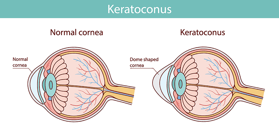
In people without keratoconus, the cornea remains smooth and rounded, like a small dome. This allows light to enter the person’s eye and focus correctly on the retina. In people with keratoconus, the cornea gets progressively weaker and thinner. As the cornea thins, it starts to bulge outward, changing the way light enters the eye.
The exact causes of keratoconus are not known. However, in the United States, it’s believed that approximately 55 out of 100,000 people have the condition, according to the American Academy of Ophthalmology. Additionally, research suggests that nearly 1 in 10 people who have keratoconus have a parent with the condition. Keratoconus usually becomes noticeable in the teenage years or early 20s, and consequently, that’s typically when it’s diagnosed.
Environmental factors that may contribute to the development of the condition include allergies and excessive eye rubbing. It’s also believed that having a connective tissue disorder, especially Marfan syndrome or Ehlers-Danlos syndrome, is associated with an increased risk.
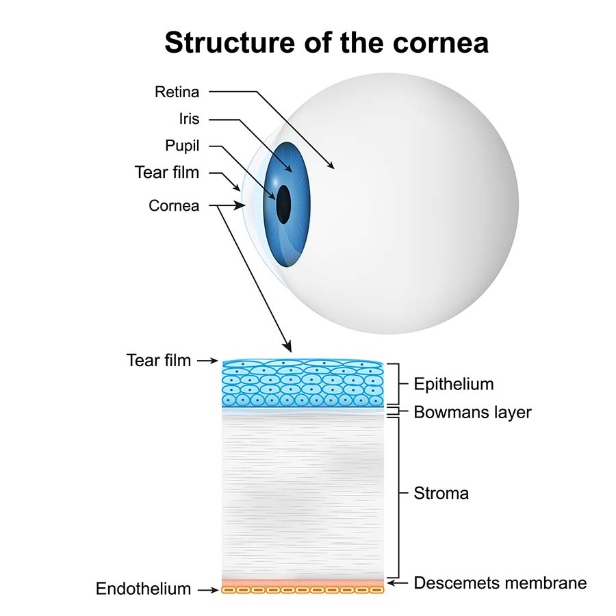
The cornea has several layers, including the epithelium, Bowman’s layer, stroma, Descemet’s membrane, and endothelium. Of those layers, the stroma is the thickest, and it’s mainly composed of type I collagen, which is the most common collagen type. In fact, it’s estimated that about 90 percent of all the collagen in the human body is type I.
Normally, the collagen in the stroma is neatly organized, like a mesh net or scaffold, where every fiber is organized and linked. In an eye that has keratoconus, the interlinked collagen scaffolding becomes weak and disorganized. Once this happens, the cornea starts to slowly bulge.
Keratoconus is a slowly progressive eye disease. People don’t often realize they have it until their glasses suddenly stop correcting their vision or their contacts no longer fit comfortably.
Keratoconus symptoms include:
Keratoconus causes a pronounced bulge in the cornea that affects the way light enters the eye. It’s important to note that keratoconus often occurs in both eyes, but one eye may be worse than the other. Because of the way keratoconus distorts the shape of the eye, it can be increasingly difficult to correct refractive errors with glasses and standard contact lenses.
Cases of astigmatism are diagnosed when the cornea is misshapen. Instead of being round, like a basketball or dome, it’s more oval-shaped, like a football. Astigmatism also doesn’t cause corneal weakness. With keratoconus, the cornea weakens, bulges, and eventually resembles a cone.
It’s important to understand that keratoconus is a progressive eye disease. This means that the condition often starts out mild, and glasses and standard contact lenses usually correct the refractive errors. However, as the disease progresses, it can be harder to correct the person’s vision. The good news is that early treatment can help preserve vision for longer.
Keratoconus doesn’t often lead to complete blindness. However, it has the potential to result in severe vision impairment. Without treatment, even specialty contact lenses, like scleral lenses, may not be able to restore clear vision.
The cornea can become too thin for treatment, which is when keratoconus is often designated as severe. At this stage, the cornea is often severely scarred, and the affected person usually has significant visual impairment. Ophthalmologists often recommend corneal transplants to help restore vision.
There are several treatments that can help treat, slow, or stop the progression of keratoconus, including scleral contact lenses and keratoconus surgery, which includes a corneal cross-linking procedure and corneal transplants.
Scleral contacts are rigid gas-permeable lenses that are designed to go completely over the cornea with the edges resting on the white part of the eye (sclera). They are often prescribed to keratoconus patients because they can help dramatically improve vision by correcting irregular astigmatism.
If you’re experiencing symptoms of keratoconus, like halos, glare or frequent changes in your glasses or contact lens prescription, you may have keratoconus. The good news is that early detection and keratoconus treatment can help slow or stop the progression. To schedule an appointment for a corneal evaluation at one of our Atlanta Eye Care Centers, contact us today.
David J. Sackel, M.D. is a board-certified ophthalmologist specializing in cornea and external disease, cataract surgery, and refractive surgery. He graduated magna cum laude from Duke University, earned his M.D. from the University of Pennsylvania School of Medicine and an MBA from the Wharton School, then completed his ophthalmology residency at New York University and a cornea fellowship at Emory University.